Be careful what you measure: Comparing measures of integrated information

entropycover2019

Our new paper on ‘measuring integrated information’ is out now, open access, in the journal Entropy. It’s part of a special issue dedicated to integrated information theory.

In consciousness research, ‘integrated information theory’, or IIT, has come to occupy a highly influential and rather controversial position. Acclaimed by some as the most important development in consciousness science so far, critiqued by others as too mathematically abstruse and empirically untestable, IIT is by turns both fascinating and frustrating. Certainly, a key challenge for IIT is to develop measures of ‘integrated information’ that can be usefully applied to actual data. These measures should capture, in empirically interesting and theoretically profound ways, the extent to which ‘a system generates more information than the sum of its parts’. Such measures are also of interest in many domains beyond consciousness, through for example to physics and engineering, where notions of ‘dynamical complexity’ are of more general importance.

Adam Barrett and I have been working towards this challenge for many years, both through approximations of the measure F (‘phi’, central to the various iterations of IIT) and through alternative measures like ‘causal density’. Alongside new work from other groups, there now exist a range of measures of integrated information – yet so far no systematic comparison of how they perform on non-trivial systems.

This is what we provide in our new paper, led by Adam along with Pedro Mediano from Imperial College London.

*

We describe, using a uniform notation, six different candidate measures of integrated information (among which we count the related measure of ‘causal density’). We set out the intuitions behind each, and compare their properties across a series of criteria. We then explore how they behave on a variety of network models, some very simple, others a little bit more complex.

The most striking finding is that the measures all behave very differently – no two measures show consistent agreement across all our analyses. Here’s an example:

screen shot 2019-01-03 at 16.45.52

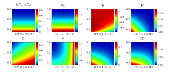
Diverse behavior of measures of integrated information. The six measures (plus two control measures) are shown in terms of their behavior on a simple 2-node network animated by autoregressive dynamics.

At first glance this seems worrying for IIT since, ideally, one would want conceptually similar measures to behave in similar ways when applied to empirical test-cases. Indeed, it is worrying if existing measures are used uncritically. However, by rigorously comparing these measures we are able to identify those which better reflect the underlying intuitions of ‘integrated information’, which we believe will be of some help as these measures continue to be developed and refined.

Integrated information, along with related notions of dynamical complexity and emergence, are likely to be important pillars of our emerging understanding of complex dynamics in all sorts of situations – in consciousness research, in neuroscience more generally, and beyond biology altogether. Our new paper provides a firm foundation for the future development of this critical line of research.

*

One important caveat is necessary. We focus on measures that are, by construction, applicable to the empirical, or spontaneous, statistically stationary distribution of a system’s dynamics. This means we depart, by necessity, from the supposedly more fundamental measures of integrated information that feature in the most recent iterations of IIT. These recent versions of the theory appeal to the so-called ‘maximum entropy’ distribution since they are more interested in characterizing the ‘cause-effect structure’ of a system than in saying things about its dynamics. This means we should be very cautious about taking our results to apply to current versions of IIT. But, in recognizing this, we also return to where we started in this post. A major issue for the more recent (and supposedly more fundamental) versions of IIT is that they are extremely challenging to operationalize and therefore to put to an empirical test. Our work on integrated information departs from ‘fundamental’ IIT precisely because we prioritise empirical applicability. This, we think, is a feature, not a bug.

*

All credit for this study to Pedro Mediano and Adam Barrett, who did all the work. As always, I’d like to thank the Dr. Mortimer and Theresa Sackler Foundation, and the Canadian Institute for Advanced Research, Azrieli Programme in Brain, Mind, and Consciousness, for their support. The paper was published in Entropy on Christmas Day, which may explain why some of you might’ve missed it!  But it did make the cover, which is nice.

*

Mediano, P.A.M., Seth, A.K., and Barrett, A.B. (2019). Measuring integrated information. Comparison of candidate measures in theory and in simulation. Entropy, 21:17

Can we figure out the brain’s wiring diagram?

connecttomemain_2

The human brain, it is often said, is the most complex object in the known universe. Counting all the connections among its roughly 90 billion neurons, at the rate of one each second, would take about 3 million years – and just counting these connections says nothing about their intricate patterns of connectivity. A new study, published this week in Proceedings of the National Academy of Sciences USA, shows that mapping out these patterns is likely to be much more difficult than previously thought — but also shows what we need to do, to succeed.

Characterizing the detailed point-to-point connectivity of the brain is increasingly recognized as a key objective for neuroscience. Many even think that without knowing the ‘connectome’ – the brain’s wiring diagram – we will never understand how its electrochemical alchemy gives rise to our thoughts, actions, perceptions, beliefs, and ultimately to our consciousness. There is a good precedent for thinking along these lines. Biology has been galvanized by sequencing of the genome (of humans and of other species), and genetic medicine is gathering pace as whole-genome sequencing becomes fast and cheap enough to be available to the many, not just the few. Big-science big-money projects like the Human Genome Project were critical to these developments. Similar efforts in brain science – like the Human Connectome Project in the US and the Human Brain Project in Europe – are now receiving vast amounts of funding (though not without criticism, especially in the European case) (see also here). The hope is that the genetic revolution can be replicated in neuroscience, delivering step changes in our understanding of the brain and in our ability to treat neurological and psychiatric disorders.

Mapping the networks of the human brain relies on non-invasive neuroimaging methods that can be applied without risk to living people. These methods almost exclusively depend on ‘diffusion magnetic resonance imaging (dMRI) tractography’. This technology measures, for each location (or ‘voxel’) in the brain, the direction in which water is best able to diffuse. Taking advantage of the fact that water diffuses more easily along the fibre bundles connecting different brain regions, than across them, dMRI tractography has been able to generate accurate, informative, and surprisingly beautiful pictures of the major superhighways in the brain.

Diffusion MRI of the human brain.  Source: Human Connectome Project.

Diffusion MRI of the human brain. Source: Human Connectome Project.

But identifying these neuronal superhighways is only a step towards the connectome. Think of a road atlas: knowing only about motorways may tell you how cities are connected, but its not going to tell you how to get from one particular house to another. The assumption in neuroscience has been that as brain scanning improves in resolution and as tracking algorithms gain sophistication, dMRI tractography will be able to reveal the point-to-point long-range anatomical connectivity needed to construct the full connectome.

In a study published this week we challenge this assumption, showing that basic features of brain anatomy pose severe obstacles to measuring cortical connectivity using dMRI. The study, a collaboration between the University of Sussex in the UK and the National Institutes of Health (NIH) in the US, applied dMRI tractography to ultra-high resolution dMRI data obtained from extensive scanning of the macaque monkey brain – data of much higher quality than can be presently obtained from human studies. Our analysis, led by Profs. Frank Ye and David Leopold of NIH and Ph.D student Colin Reveley of Sussex, took a large number of starting points (‘seed voxels’) in the brain, and investigated which other parts of the brain could be reached using dMRI tractography.

The result: roughly half of the brain could not be reached, meaning that even our best methods for mapping the connectome aren’t up to the job. What’s more, by looking carefully at the actual brain tissue where tractography failed, we were able to figure out why. Lying just beneath many of the deep valleys in the brain (the ‘sulci’ – but in some other places too), are dense weaves of neuronal fibres (‘white matter’) running largely parallel to the cortical surface. The existence of these ‘superficial white matter fibre systems’, as we call them, prevents the tractography algorithms from detecting where small tributaries leave the main neuronal superhighways, cross into the cortical grey matter, and reach their destinations. Back to the roads: imagine that small minor roads occasionally leave the main motorways, which are flanked by other major roads busy with heavy traffic. If we tried to construct a detailed road atlas by measuring the flow of vehicles, we might well miss these small but critical branching points.

This image shows, on a colour scale, the 'reachability' of different parts of the brain by diffusion tractography.

This image shows, on a colour scale, the ‘reachability’ of different parts of the brain by diffusion tractography.

Identifying the connectome remains a central objective for neuroscience, and non-invasive brain imaging – especially dMRI – is a powerful technology that is improving all the time. But a comprehensive and accurate map of brain connectivity is going to require more than simply ramping up scanning resolution and computational oomph, a message that mega-budget neuroscience might usefully heed. This is not bad news for brain research. Solving a problem always requires fully understanding what the problem is, and our findings open new opportunities and objectives for studies of brain connectivity. Still, it goes to show that the most complex object in the universe is not quite ready to give up all its secrets.


Colin Reveley, Anil K. Seth, Carlo Pierpaoli, Afonso C. Silva, David Yu, Richard C. Saunders, David A. Leopold*, and Frank Q. Ye. (2015) Superficial white-matter fiber systems impede detection of long-range cortical connections in diffusion MR tractography. Proc. Nat. Acad. Sci USA (2015). doi/10.1073/pnas.1418198112

*David A. Leopold is the corresponding author.

The Human Brain Project risks becoming a missed opportunity

Image concept of a network of neurons in the human brain.

The brain is much on our minds at the moment. David Cameron is advocating a step-change in dementia research, brain-computer interfaces promise new solutions to paralysis, and the ongoing plight of Michael Schumacher has reminded us of the terrifying consequences of traumatic brain injury. Articles in scholarly journals and in the media are decorated with magical images of the living brain, like the one shown below, to illuminate these stories. Yet, when asked, most neuroscientists will say we still know very little about how the brain works, or how to fix it when it goes wrong.

DTI-sagittal-fibers
A diffusion tensor image showing some of the main pathways along which brain connections are organized.

The €1.2bn Human Brain Project (HBP) is supposed to change all this. Funded by the European Research Council, the HBP brings together more than 80 research institutes in a ten-year endeavour to unravel the mysteries of the brain, and to emulate its powers in new technologies. Following examples like the Human Genome Project and the Large Hadron Collider (where Higgs’ elusive boson was finally found), the idea is that a very large investment will deliver very significant results. But now a large contingent of prominent European neuroscientists are rebelling against the HBP, claiming that its approach is doomed to fail and will undermine European neuroscience for decades to come.

Stepping back from the fuss, it’s worth thinking whether the aims of the HBP really make sense. Sequencing the genome and looking for Higgs were both major challenges, but in these cases the scientific community agreed on the objectives, and on what would constitute success. There is no similar consensus among neuroscientists.

It is often said that the adult human brain is the most complex object in the universe. It contains about 90 billion neurons and a thousand times more connections, so that if you counted one connection each second it would take about three million years to finish. The challenge for neuroscience is to understand how this vast, complex, and always changing network gives rise to our sensations, perceptions, thoughts, actions, beliefs, desires, our sense of self and of others, our emotions and moods, and all else that guides our behaviour and populates our mental life, in health and in disease. No single breakthrough could ever mark success across such a wide range of important problems.

The central pillar of the HBP approach is to build computational simulations of the brain. Befitting the huge investment, these simulations would be of unprecedented size and detail, and would allow brain scientists to integrate their individual findings into a collective resource. What distinguishes the HBP – besides the money – is its aggressively ‘bottom up’ approach: the vision is that by taking care of the neurons, the big things – thoughts, perceptions, beliefs, and the like – will take care of themselves. As such, the HBP does not set out to test any specific hypothesis or collection of hypotheses, marking another distinction with common scientific practice.

Could this work? Certainly, modern neuroscience is generating an accelerating data deluge demanding new technologies for visualisation and analysis. This is the ‘big data’ challenge now common in many settings. It is also clear that better pictures of the brain’s wiring diagram (the ‘connectome’) will be essential as we move ahead. On the other hand, more detailed simulations don’t inevitably lead to better understanding. Strikingly, we don’t fully understand the brain of the tiny worm Caenorhabtis elegans even though it has only 302 neurons and the wiring diagram is known exactly. More generally, a key ability in science is to abstract away from the specifics to see more clearly what underlying principles are at work. In the limit, a perfectly accurate model of the brain may become as difficult to understand as the brain itself, as Borges long ago noted when describing the tragic uselessness of the perfectly detailed map.

jorge_luis_borges_por_paola_agosti
Jorge Luis Borges at Harvard University, 1967/8

Neuroscience is, and should remain, a broad church. Understanding the brain does not reduce to simulating the collective behaviour of all its miniscule parts, however interesting a part of the final story this might become. Understanding the brain means grasping complex interactions cross-linking many different levels of description, from neurons to brain regions to individuals to societies. It means complementing bottom-up simulations with new theories describing what the brain is actually doing, when its neurons are buzzing merrily away. It means designing elegant experiments that reveal how the mind constructs its reality, without always worrying about the neuronal hardware underneath. Sometimes, it means aiming directly for new treatments for devastating neurological and psychiatric conditions like coma, paralysis, dementia, and depression.

Put this way, neuroscience has enormous potential to benefit society, well deserving of high profile and large-scale support. It would be a great shame if the Human Brain Project, through its singular emphasis on massive computer simulation, ends up as a lightning rod for dissatisfaction with ‘big science’ rather than fostering a new and powerfully productive picture of the biological basis of the mind.

This article first appeared online in The Guardian on July 8 2014.  It appeared in print in the July 9 edition, on page 30 (comment section).

Post publication notes:

The HBP leadership have published a response to the open letter here. I didn’t find it very convincing. There have been a plethora of other commentaries on the HBP, as it comes up to its first review.  I can’t provide an exhaustive list but I particularly liked Gary Marcus’ piece in the New York Times (July 11). There was also trenchant criticism in the editorial pages of Nature.  Paul Verschure has a nice TED talk addressing some of the challenges facing big data, encompassing the HBP.


How does the brain fit into the skull?

Image

Announcing a new paper co-authored with David Samu and Thomas Nowotny, published yesterday in the open-access journal PLoS Computational Biology.

Influence of Wiring Cost on the Large-Scale Architecture of Human Cortical Connectivity

Macroscopic regions in the grey matter of the human brain are intricately connected by white-matter pathways, forming the extremely complex network of the brain. Analysing this brain network may provide us insights on how anatomy enables brain function and, ultimately, cognition and consciousness. Various important principles of organization have indeed been consistently identified in the brain’s structural connectivity, such as a small-world and modular architecture. However, it is currently unclear which of these principles are functionally relevant, and which are merely the consequence of more basic constraints of the brain, such as its three-dimensional spatial embedding into the limited volume of the skull or the high metabolic cost of long-range connections. In this paper, we model what aspects of the structural organization of the brain are affected by its wiring constraints by assessing how far these aspects are preserved in brain-like networks with varying spatial wiring constraints. We find that all investigated features of brain organization also appear in spatially constrained networks, but we also discover that several of the features are more pronounced in the brain than its wiring constraints alone would necessitate. These findings suggest the functional relevance of the ‘over-expressed’ properties of brain architecture.

New: Image from this paper featured as MRC biomedical image of the day on April 29th 2014!

brainteaser